先に結論:乳腺MRIのassociated features(随伴所見)
- ACR BI-RADS Atlas 第5版(2013)のMRI用語では、所見をfocus・mass(腫瘤)・non-mass enhancement(NME、非腫瘤性増強)に分け、ほかにnon-enhancing findings(造影されない所見)・associated features(随伴所見)・fat-containing lesions(脂肪を含む病変)などの項目で記載します(Choi 2023)
- 第5版のassociated featuresは、乳頭陥凹・乳頭浸潤・皮膚陥凹・皮膚肥厚・皮膚浸潤(直接浸潤、炎症性乳癌)・腋窩リンパ節腫大・大胸筋浸潤・胸壁浸潤・構築の乱れです(Choi 2023)
- 以前は随伴所見として並べられることのあった造影前T1強調像での乳管内高信号・嚢胞・術後の血腫/漿液腫・異物による信号欠損は、第5版ではnon-enhancing findingsの側に整理されています(Choi 2023)
- これらは悪性を示唆したり、病期(T因子・N因子)の判断に関わるため、T2強調像・造影前T1強調像もあわせて評価し、レポートに記載します
- 2025年のBI-RADS v2025では、associated featuresのinvasion(浸潤)がinvolvementに言い換えられ、peritumoral edema(腫瘍周囲の浮腫)が新しい随伴所見に加わり、腋窩リンパ節腫大はリンパ節の独立した項目に移り、構築の乱れはこの項目から外れました(Kim 2026)
associated features(第5版の随伴所見)
以下のような所見があればT2強調像やT1強調像なども併せて評価をして、記載する。第4版までの文献や旧来の報告書では associated findings と書かれていることもあります。
Associated features(BI-RADS 第5版, 2013)
- nipple retraction 乳頭陥凹
- nipple invasion 乳頭浸潤
- skin retraction 皮膚陥凹
- skin thickening 皮膚肥厚
- skin invasion(direct invasion / inflammatory cancer) 皮膚浸潤(直接浸潤・炎症性乳癌)
- axillary adenopathy 腋窩リンパ節腫大
- pectoralis muscle invasion 大胸筋浸潤
- chest wall invasion 胸壁浸潤
- architectural distortion 構築の乱れ
(Choi 2023の第5版MRI用語表による)
旧来の一覧の項目は第5版でどこに入ったか
| 旧来の一覧にあった項目 | 第5版での扱い |
|---|---|
| high signal in duct on pre-contrast scan(造影前の乳管内高信号) | non-enhancing findings:ductal precontrast high signal on T1W |
| cysts(嚢胞) | non-enhancing findings:cyst |
| hematoma/blood(血腫/血液) | non-enhancing findings:postoperative collections(hematoma/seroma) |
| abnormal signal void(異常信号欠損) | non-enhancing findings:signal void from foreign bodies, clips など |
| lymphadenopathy(リンパ節腫脹) | associated features:axillary adenopathy |
| chest wall or skin invasion(胸壁・皮膚浸潤) | associated features:chest wall invasion / pectoralis muscle invasion / skin invasion に分けて記載 |
| architectural distortion(構築の乱れ) | associated features と non-enhancing findings の両方に項目がある |
| edema(浮腫)・fluid-filled dilated ducts(拡張乳管内液体貯留) | 第5版の用語表(Choi 2023)には独立した項目としては見当たらない。治療後の皮膚・梁柱の肥厚は non-enhancing findings に入る |
non-enhancing findings には、ほかにnon-enhancing mass(造影されない腫瘤)やpost-therapy skin and trabecular thickening(治療後の皮膚・梁柱の肥厚)も含まれます。脂肪を含む病変(リンパ節、脂肪壊死、過誤腫、脂肪を含む術後の漿液腫・血腫)は fat-containing lesions として別に扱います(Choi 2023)。
症例 30歳代女性(右炎症性乳癌)

引用:Radiopaedia(Inflammatory breast cancer)
術前化学療法の前に撮影された造影脂肪抑制T1強調像の横断像です(画像の左が右乳房)。右乳房に辺縁不整・スピキュラを伴う腫瘤があり、皮膚は著明に肥厚して強く造影され、右腋窩には壊死を伴うリンパ節が集簇して腫大しています。この症例では乳頭浸潤・皮膚浸潤・大胸筋浸潤・腋窩リンパ節腫大と、随伴所見が多く報告されています。
腫瘤・非腫瘤性増強・造影パターンの用語(第5版)
| 項目 | 第5版の用語 |
|---|---|
| 乳腺の背景 | 乳腺量(amount of fibroglandular tissue):a〜d の4段階。背景乳腺増強(background parenchymal enhancement:BPE):minimal・mild・moderate・marked、symmetric・asymmetric |
| 腫瘤の形 | oval(lobulatedを含む)・round・irregular |
| 腫瘤の辺縁 | circumscribed / not circumscribed(irregular・spiculated)。第4版の smooth は circumscribed に変更 |
| 腫瘤の内部造影 | homogeneous・heterogeneous・rim enhancement・dark internal septations(造影されない隔壁) |
| 非腫瘤性増強の分布 | focal・linear・segmental・regional・multiple regions・diffuse。ductal は第5版で削除 |
| 非腫瘤性増強の内部パターン | homogeneous・heterogeneous・clumped・clustered ring(第5版で追加)。reticular/dendritic・stippled/punctate は削除 |
| 造影の時間変化(kinetic curve assessment) | initial phase(早期相):slow・medium・fast。delayed phase(後期相):persistent・plateau・washout |
(Choi 2023;Lunkiewicz 2020;Kaplan 2026)
dark internal septationsは線維腺腫に特異的とされ、spiculatedな辺縁やrim enhancementは悪性を示唆します(Nunes 1997)。266病変の検討では、乳癌の57.4%がwashout型、良性の83.0%がpersistent型でした(Kuhl 1999)。
第5版で加わった clustered ring と非腫瘤性増強の見方
- clustered ring は、乳管周囲の間質が細い輪状に造影されて集まって見えるパターンで、第5版で非腫瘤性増強の内部パターンに加わりました。生検を行ったNME 72件の検討では、clustered ringの悪性の陽性的中率は53.85%、segmental分布は62.5%で、両者がそろうと66.67%でした(Lunkiewicz 2020)。
- 一方、NMEとして報告された386所見を第5版で見直した検討では、20.0%がBPE(背景乳腺増強)と判断し直されました。陽性的中率はclustered ringで36.7%、clumpedで27.5%、分布ではsegmentalで34.5%で、悪性NMEの3分の1はT2強調像で高信号でした(Chikarmane 2017)。T2高信号だけで良性と考えないことが大切です。
- BI-RADS 3と報告された280所見を3人で読み直した検討では、腫瘤の形・辺縁・内部造影やNMEの分布・内部パターンの一致度は中等度〜高い一方、kineticの一致度は低いという結果でした(Grimm 2015)。
参考:BI-RADS v2025での変更
2013年の第5版から12年後に出た ACR BI-RADS v2025 では、MRIの kinetic の「initial phase」が「early phase」に改められ、腫瘤の形に lobulated が加わり、腫瘤のT2信号(hyperintense / not hyperintense)が新しい項目として入りました。symmetric / asymmetric はNMEの分布から外れてBPEの側に整理されています(Kaplan 2026)。このページの用語は第5版(2013)に合わせ、v2025での違いは必要な箇所で補っています。
BI-RADS v2025(2025年)での随伴所見の変更点
2013年の第5版から12年ぶりに改訂されたBI-RADS v2025は、診断の考え方を大きく変えるものではなく、用語の整理と構成の見直しが中心とされています(Kim 2026)。随伴所見に関する主な変更は次のとおりです。
| 第5版(2013年) | v2025(2025年) | 変更 |
| nipple invasion(乳頭浸潤) | nipple involvement | invasionがinvolvementに |
| skin invasion(皮膚浸潤) | skin involvement | invasionがinvolvementに |
| pectoralis muscle invasion(大胸筋浸潤) | pectoralis muscle involvement | invasionがinvolvementに |
| chest wall invasion(胸壁浸潤) | chest wall involvement | invasionがinvolvementに |
| axillary adenopathy(腋窩リンパ節腫大) | リンパ節の独立した項目へ | 随伴所見から削除 |
| architectural distortion(構築の乱れ) | なし | 削除 |
| なし | peritumoral edema(腫瘍周囲の浮腫) | 追加 |
| nipple retraction・skin retraction・skin thickening | 同じ | 変更なし |
(Kim 2026の表をもとに作成)
peritumoral edema(腫瘍周囲の浮腫)
- 悪性または疑わしい病変の周囲の乳腺に見られる、T2強調像での高信号と定義されています(Kim 2026)。
- 悪性を示唆する所見で、広範囲に病変がある患者ではリンパ節転移のリスクが高いことと関連していました。一方で、生検後の変化など良性の状態でも見られます(Kim 2026)。T2強調像の浮腫だけで悪性とは決められません。生検の既往を確認します。
リンパ節は独立した項目に
- v2025では、リンパ節は随伴所見ではなく独立した項目になり、乳房内・腋窩・内胸の3か所について、それぞれ正常か異常かで記載します(Kim 2026)。
- 乳房MRIでのリンパ節評価について、妥当性が確認されたサイズや皮質の厚さの基準値はなく、形態、以前の画像や反対側のリンパ節との比較、臨床の状況で判断するとされています(Kim 2026)。
造影されない所見・その他
- 第5版のnon-enhancing findingsは、v2025ではOther Findings—Typically Benign(通常は良性のその他の所見)に改められ、fat-containing lesions(脂肪を含む病変)の項目は廃止されて、個々の所見が他の項目に振り分けられました(Kim 2026)。
インプラント(breast implants)の用語
- 第5版で整備されたインプラントの用語(retroglandular・retropectoralなどの位置、focal bulge、radial folds、subcapsular line、keyhole sign、linguine sign、被膜外のシリコーン、water droplets、peri-implant fluid)は、v2025でも言葉は変わらず、図が更新され、簡略化したインプラント用MRIプロトコルの案内が加わりました(Kaplan 2026)。
引用元の図 インプラントの位置(retroglandular・retropectoral)
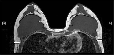
両側のシリコーンゲル充填インプラントが大胸筋より前方にあり、retroglandular(乳腺の後ろ)の位置にあることを示すT1強調像の横断像です。矢印は大胸筋です。
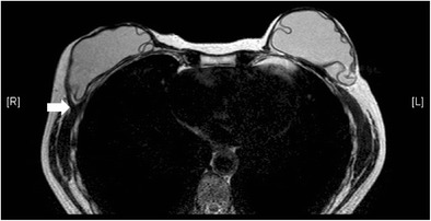
右のシリコーンゲル充填インプラントがretropectoral(大胸筋の後ろ)の位置にある、T2強調像の横断像です。右の大胸筋(矢印)がインプラントで分けられています。
引用:Wong T et al. Insights Imaging 2016(Figure 1・Figure 2)(CC BY 4.0)
あわせて読みたい:乳腺MRIのBI-RADSカテゴリー分類と読影法 / 非腫瘤性増強(NME)の読影法
乳腺MRIの随伴所見でよくある疑問(Q&A)
Q1. v2025で、浸潤の「invasion」が「involvement」になったのはなぜですか。
乳頭・皮膚・大胸筋・胸壁の所見の用語が、invasionからinvolvementに改められました(Kim 2026)。改訂の理由の詳しい説明は、BI-RADS v2025の原典で確認してください。
Q2. 腋窩リンパ節腫大は、どこに書きますか。
v2025では随伴所見ではなく、リンパ節の項目に、乳房内・腋窩・内胸ごとに正常か異常かで記載します(Kim 2026)。
Q3. 腫瘍の周りのT2高信号は、すべて悪性を意味しますか。
いいえ。peritumoral edemaは悪性を示唆する所見ですが、生検後の変化など良性でも見られます(Kim 2026)。
※乳房のしこり、皮膚の赤み・ひきつれ、乳頭の変化など気になる症状があるときは、医療機関(乳腺外科など)を受診してください。
参考文献
- Shin K, Phalak K, Hamame A, et al. Interpretation of Breast MRI Utilizing the BI-RADS Fifth Edition Lexicon: How Are We Doing and Where Are We Headed? Curr Probl Diagn Radiol. 2017;46(1):26-34. PubMed
- Spak DA, Plaxco JS, Santiago L, et al. BI-RADS fifth edition: A summary of changes. Diagn Interv Imaging. 2017;98(3):179-190. PubMed
- Rao AA, Feneis J, Lalonde C, et al. A Pictorial Review of Changes in the BI-RADS Fifth Edition. Radiographics. 2016;36(3):623-39. PubMed
- Griffith BM, Taylor CR, Hawley JR, et al. Applying the Contrast-enhanced Mammography BI-RADS Lexicon to Clinical Practice. Radiographics. 2025;45(11):e240136. PubMed
- Nunes LW, Schnall MD, Siegelman ES, et al. Diagnostic performance characteristics of architectural features revealed by high spatial-resolution MR imaging of the breast. AJR Am J Roentgenol. 1997;169(2):409-15. PubMed
- Kuhl CK, Mielcareck P, Klaschik S, et al. Dynamic breast MR imaging: are signal intensity time course data useful for differential diagnosis of enhancing lesions? Radiology. 1999;211(1):101-10. PubMed
- Choi JS. Breast Imaging Reporting and Data System (BI-RADS): Advantages and Limitations. J Korean Soc Radiol. 2023;84(1):3-14. PubMed
- Lunkiewicz M, Forte S, Freiwald B, et al. Interobserver variability and likelihood of malignancy for fifth edition BI-RADS MRI descriptors in non-mass breast lesions. Eur Radiol. 2020;30(1):77-86. PubMed
- Chikarmane SA, Michaels AY, Giess CS. Revisiting Nonmass Enhancement in Breast MRI: Analysis of Outcomes and Follow-Up Using the Updated BI-RADS Atlas. AJR Am J Roentgenol. 2017;209(5):1178-1184. PubMed
- Grimm LJ, Anderson AL, Baker JA, et al. Interobserver Variability Between Breast Imagers Using the Fifth Edition of the BI-RADS MRI Lexicon. AJR Am J Roentgenol. 2015;204(5):1120-4. PubMed
- Kaplan E, Aydemir AB. How Does ACR BI-RADS v2025 Change the Radiologist’s Approach? A Practical Guide Across Mammography, Ultrasound, and MRI: A Narrative Review. Diagnostics (Basel). 2026;16(13):2135. PubMed
- Kim JY. Breast Imaging Reporting and Data System v2025: Key Updates in MRI. J Korean Soc Radiol. 2026;87(3):473-484. PubMed
画像診断を無料で学べるコンテンツ

腹部画像診断を症例で学ぶ
4日に1回、朝6時に症例が届き、実際に画像をスクロールして読影する講座です。90症例以上・1年以上続く講座で、医師・医学生・放射線技師・看護師など10,000名以上が参加中です。
講座に参加する(無料) →
