先に結論:腸管壁の脂肪(fat halo sign)は正常のことも多い
- fat halo signは、腸の壁の真ん中の層(粘膜下層)に脂肪がたまり、CTで3層の的(まと)のように見える所見です
- クローン病・潰瘍性大腸炎などの慢性的な炎症、放射線腸炎、化学療法後で見られますが、病気のない人(特に肥満の人)にも正常変異として見られます
- 真ん中の層が脂肪でなく水の濃さ(water halo)なら、急性の炎症・虚血・むくみを考えます。fat halo signだけで炎症性腸疾患と決めつけないことが大切です
- target sign(ターゲットサイン)は「的のように見える」所見の総称で、腸管壁のCT所見のほか、腸重積の超音波像や神経鞘腫瘍のMRIなどでも使われます
fat halo sign / target fat patternとは
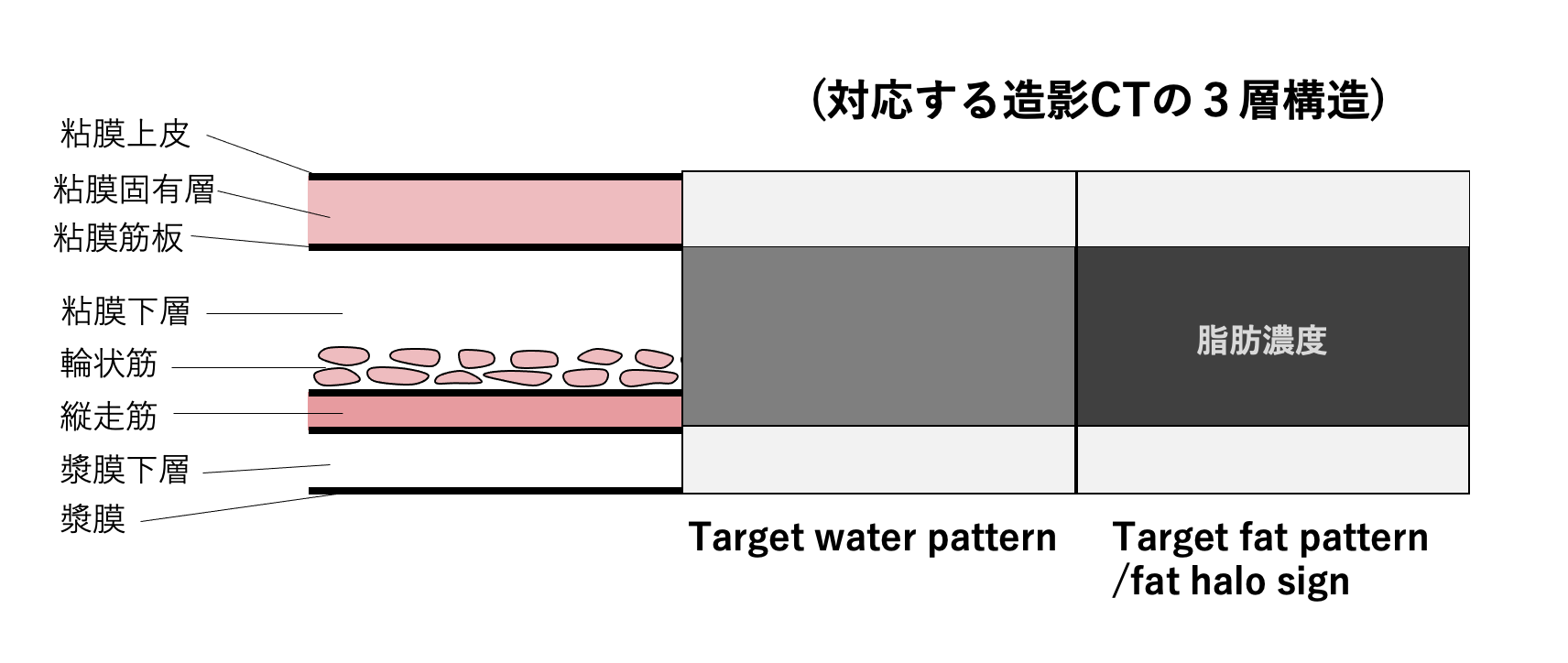
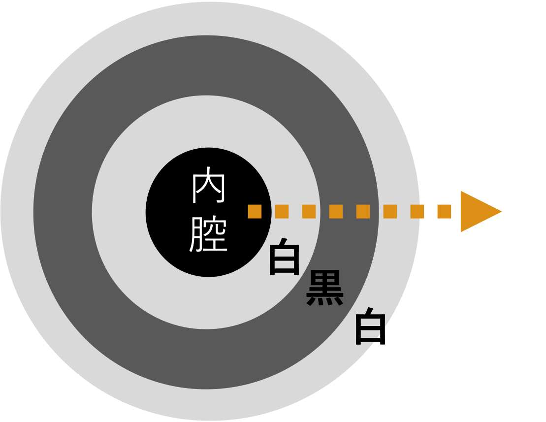
- 腹部CTでみられる fat halo sign は、肥厚した腸管壁のうち中間層(主に粘膜下層)に脂肪濃度が沈着し、内側の粘膜層と外側の筋層・漿膜側が軟部濃度のまま保たれることで、三層性のtarget外観を示す所見である。
- 文献上は「fat halo sign」「submucosal fat deposition」「fat density target sign」などの名称で記載される。
- 実際の読影では、造影CTで内側の粘膜がやや増強し、中央の粘膜下層が低吸収、外層が相対的に高吸収にみえるため、「target sign」に見える。そのうち中央層が水濃度ではなく脂肪濃度であるものを、water halo signではなくfat halo signと呼び分けるのが重要である。
- 原因としてはCrohn病、潰瘍性大腸炎など炎症性腸疾患、慢性炎症、放射線性腸炎、白血病やリンパ腫に対する化学療法後(比較的短期間で粘膜下脂肪沈着が出現しうることが報告されている)の他、消化管疾患のない人にも見られ肥満に関連した正常変異の可能性が示唆されている。
- 従って、fat halo sign単独で炎症性腸疾患(IBD)を強く疑うべきではない。腸疾患と無関係な理由(尿路結石疑い)でCTを撮影した連続100例の検討では21%にfat halo signがみられ、いずれもIBDの既往はなく、体重との有意な関連(21例中16例が90 kg超)が示されている。むしろ偶発的な壁内脂肪化として扱うほうが妥当な場面がある。同様のfat halo signは胃壁にもみられることがある。腸疾患のない胃壁fat halo sign例90例と対照130例の前向き検討では、BMIに有意差はなかった一方、内臓脂肪面積が有意に大きく、大腸・回腸のfat halo signを伴う頻度も高かったことから、体重よりも内臓脂肪量との関連が示唆されている。
症例 70歳代男性 スクリーニング(既往歴なし)
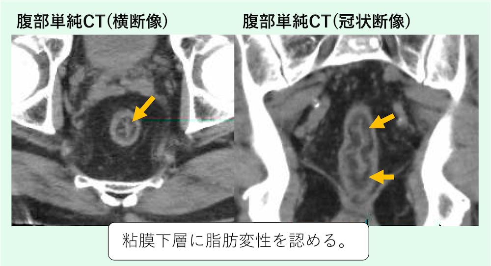
直腸の粘膜下層に3層構造を認め、粘膜下層は脂肪濃度(fat halo sign / target fat pattern)となっています。
既往歴はなく、生理的な正常変異と考えられます。
target sign(ターゲットサイン)とは?検査ごとの意味の違い
target signは同心円状に層が重なって的のように見える所見の呼び名で、検査や臓器によって意味が変わります。
| 場面 | 見え方 | 主に考えるもの |
|---|---|---|
| CT:腸管壁(water halo sign) | 肥厚した壁が2〜3層に分かれ、中間層が水の濃さ | 炎症性腸疾患・血管性疾患・感染・放射線障害(Wittenberg 2002) |
| CT:腸管壁(fat halo sign) | 3層のうち中間層(粘膜下層)が脂肪の濃さ | 小腸のCrohn病、大腸の炎症性腸疾患、正常変異(Wittenberg 2002、Harisinghani 2003) |
| 超音波:腸重積 | 横断像で腸管が何重にも重なった同心円(多重同心円サイン・ドーナツ状) | 腸重積(del-Pozo 1999) |
| MRI:末梢神経鞘腫瘍 | 辺縁が高信号・中心が低信号の2層 | 良性の神経鞘腫瘍を示唆(Ahlawat 2018) |
fat halo signでよくある疑問(Q&A)
Q. 大腸脂肪腫とはどう違いますか?
A. 腸の脂肪腫は、CTで脂肪の濃さをもつかたまり(腫瘤)として見えます(Kakitsubata 1993)。fat halo signは腫瘤ではなく、腸の壁に沿って層状に脂肪が並ぶ点が異なります。
Q. 健診のCTでfat halo signと言われたら?
A. 症状がなければ正常変異のことも多い所見です。腹痛や下痢・血便が続くなど気になる症状があれば、消化器内科で相談してください。
関連動画:こちらでもtarget fat pattern(fat halo sign )について取り上げています。
関連記事:
出典
- Wittenberg J, Harisinghani MG, Jhaveri K, Varghese J, Mueller PR. Algorithmic approach to CT diagnosis of the abnormal bowel wall. Radiographics. 2002;22(5):1093-1107. doi:10.1148/rg.225015084.
- Fernandes T, Oliveira MI, Castro R, et al. Bowel wall thickening at CT: simplifying the diagnosis. Insights Imaging. 2014;5(2):195-208.
- Amitai MM, Arazi-Kleinman T, Avidan B, et al. Fat halo sign in the bowel wall of patients with Crohn’s disease. Clin Radiol. 2007;62(10):994-997. doi:10.1016/j.crad.2007.04.007.
- Harisinghani MG, Wittenberg J, Lee W, et al. Bowel wall fat halo sign in patients without intestinal disease. AJR Am J Roentgenol. 2003;181(3):781-784.
- Muldowney SM, Balfe DM, Hammerman AM, Wick MR. “Acute” fat deposition in bowel wall submucosa: CT appearance. J Comput Assist Tomogr. 1995;19(3):390-393. doi:10.1097/00004728-199505000-00010.
- Chen S, Harisinghani MG, Wittenberg J. Small bowel CT fat density target sign in chronic radiation enteritis. Australas Radiol. 2003;47(4):450-452.
- Gore RM, Marn CS, Kirby DF, et al. CT findings in ulcerative, granulomatous, and indeterminate colitis. AJR Am J Roentgenol. 1984;142(6):1173-1176.
- Klein HM, Günther RW, Fehlhaber C, et al. The computed tomographic morphology of Crohn’s disease and ulcerative colitis. Rofo. 1995;163(3):187-193.
- Byun JY, Ha HK, Yu SY, et al. CT features of systemic lupus erythematosus in patients with acute abdominal pain: emphasis on ischemic bowel disease. Radiology. 1999;211(1):203-209. doi:10.1148/radiology.211.1.r99mr17203.
- Ripollés T, Martínez-Pérez MJ, Paredes JM, et al. Submucosal fat accumulation in Crohn’s disease: evaluation with sonography. Intest Res. 2023;21(3):385-391. doi:10.5217/ir.2022.00030.
あわせて読みたい:回盲弁(バウヒン弁)の脂肪増生のCT画像所見
参考文献
- Harisinghani MG, Wittenberg J, Lee W, et al. Bowel wall fat halo sign in patients without intestinal disease. AJR Am J Roentgenol. 2003;181(3):781-784. PubMed
- Wittenberg J, Harisinghani MG, Jhaveri K, et al. Algorithmic approach to CT diagnosis of the abnormal bowel wall. Radiographics. 2002;22(5):1093-1107. PubMed
- Kupeli A, Danisan G, Kocak M, et al. Gastric wall fat halo sign in patients without intestinal disease. Clin Imaging. 2019;54:31-36. PubMed
- Kani KK, Moshiri M, Bhargava P, et al. Extrahepatic, nonneoplastic, fat-containing lesions of the abdominopelvic cavity: spectrum of lesions, significance, and typical appearance on multidetector computed tomography. Curr Probl Diagn Radiol. 2012;41(2):56-72. PubMed
- del-Pozo G, Albillos JC, Tejedor D, et al. Intussusception in children: current concepts in diagnosis and enema reduction. Radiographics. 1999;19(2):299-319. PubMed
- Ahlawat S, Fayad LM. Imaging cellularity in benign and malignant peripheral nerve sheath tumors: Utility of the “target sign” by diffusion weighted imaging. Eur J Radiol. 2018;102:195-201. PubMed
- Kakitsubata Y, Kakitsubata S, Nagatomo H, et al. CT manifestations of lipomas of the small intestine and colon. Clin Imaging. 1993;17(3):179-182. PubMed
画像診断を無料で学べるコンテンツ

無料講座
腹部画像診断を症例で学ぶ
4日に1回、朝6時に症例が届き、実際に画像をスクロールして読影する講座です。90症例以上・1年以上続く講座で、医師・医学生・放射線技師・看護師など10,000名以上が参加中です。
講座に参加する(無料) →