先に結論:側頭葉幹(temporal stem)
- 側頭葉幹は側頭葉と前頭葉・頭頂後頭葉をつなぐ白質の「茎」で、狭い場所に鉤状束・下前頭後頭束・前交連・視放線(Meyerループ)が集まっています
- そのため、腫瘍・炎症・虚血・浮腫や手術がこの部位に及ぶと、上四分盲などの視野障害や高次機能障害が出やすくなります
- 解剖の確認には冠状断像が分かりやすく、側頭葉の手術前の評価でも重要な部位です
実際の画像で確かめるには、画像診断cafeの頭部MRIの正常解剖ツールで、スライスを動かしながら構造をタップすると名前が表示されます(無料・登録不要)。全体の見方は頭部MRIの解剖にまとめています。
側頭葉幹とは?
- 側頭葉幹(temporal stem)(読み方は「そくとうようかん」)は、側頭葉と前頭葉をつなぐ白質。
- 狭小な領域に重要な線維路が密集するため、病変の波及(腫瘍・炎症・虚血・浮腫)や手術操作の影響が、視野障害・高次機能障害として出やすい部位として重要。
側頭葉幹を構成する主な線維路
側頭葉幹は「空間」として定義され、その内部を複数の重要な線維束が通過する。臨床的に特に重要なのは以下である。
- 鉤状束(uncinate fasciculus:UF):前頭葉眼窩面〜前側頭葉(扁桃体周辺を含む)を連絡
- 下前頭後頭束(inferior fronto-occipital fasciculus:IFOF):前頭葉と後方領域を連絡する長連合線維(側頭幹を通過)
- 前交連(anterior commissure):両側側頭葉を結ぶ交連線維(側頭幹近傍を走行)
- 視放線の前方(Meyer’s loop):側頭葉前方へ回り込む視放線で、損傷すると上四分盲が典型
側頭葉幹の正常解剖
- 冠状断像が正常解剖の把握に特に重要。
- 側頭葉幹は前後方向では扁桃体の高さから外側膝状体の高さまで広がり、鉤状束と下前頭後頭束はここを通って最外包・外包へ入る(Kier 2004)。
- Meyerループの前端は扁桃体の高さまで達し、鉤状束・前交連と隣り合う(Kier 2004)。Meyerループの先端から側頭極までの距離は手術患者で24〜43mm(平均34mm)と個人差が大きい(Yogarajah 2009)。
冠状断とは?MRI冠状断で見る側頭葉の解剖
- 冠状断(コロナル断)は、顔と平行な面で前から後ろへ輪切りにした断面です。
- 側頭葉は冠状断で見ると、外側から上側頭回・中側頭回・下側頭回が並び、側頭葉内側には海馬・扁桃体・海馬傍回があります。
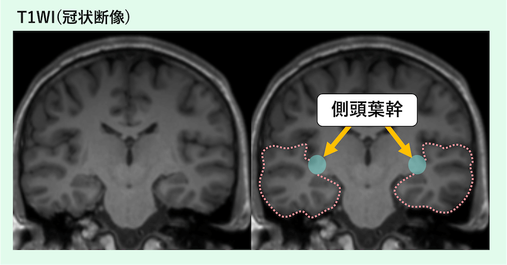
側頭葉の根や茎のように見える白質部分が側頭葉幹です。
参考文献
- Kier EL, et al. MR Imaging of the Temporal Stem: Anatomic Dissection, Tractography of the Uncinate Fasciculus, Inferior Occipitofrontal Fasciculus, and Meyer’s Loop of the Optic Radiation. AJNR (2004). PubMed
- Choi CY, et al. A Understanding of the Temporal Stem. J Korean Neurosurg Soc (2010). PubMed
- Yogarajah M, et al. Defining Meyer’s loop–temporal lobe resections, visual field deficits and diffusion tensor tractography. Brain (2009). PubMed
- Winston GP, et al. Optic Radiation Tractography and Vision in Anterior Temporal Lobe Resection. Ann Neurol (2012). PubMed
- Taoka T, et al. Diffusion Tensor Tractography of the Meyer Loop in Cases of Temporal Lobe Resection for Temporal Lobe Epilepsy. AJNR (2008). PubMed
- Peltier J, Verclytte S, Delmaire C, et al. Microsurgical anatomy of the temporal stem: clinical relevance and correlations with diffusion tensor imaging fiber tracking. J Neurosurg. 2010;112(5):1033-1038. PubMed
- Martino J, Vergani F, Robles SG, et al. New insights into the anatomic dissection of the temporal stem with special emphasis on the inferior fronto-occipital fasciculus: implications in surgical approach to left mesiotemporal and temporoinsular structures. Neurosurgery. 2010;66(3 Suppl Operative):4-12. PubMed
- Ebeling U, Reulen HJ. Neurosurgical topography of the optic radiation in the temporal lobe. Acta Neurochir (Wien). 1988;92(1-4):29-36. PubMed
画像診断を無料で学べるコンテンツ

無料講座
腹部画像診断を症例で学ぶ
4日に1回、朝6時に症例が届き、実際に画像をスクロールして読影する講座です。90症例以上・1年以上続く講座で、医師・医学生・放射線技師・看護師など10,000名以上が参加中です。
講座に参加する(無料) →