先に結論:トキソプラズマ脳症
- トキソプラズマ脳症はAIDS(CD4<100/μL)などの細胞性免疫低下で、潜伏していた原虫が再活性化して起こり、AIDS患者の脳腫瘤の代表です
- MRIでは基底核・皮髄境界の多発するリング状に造影される病変と浮腫が典型で、リングの中に偏った結節(eccentric target sign)が特徴的です
- 最大の鑑別は中枢神経原発悪性リンパ腫で、DWI・ADC・FDG-PET・タリウムSPECTなどを参考にし、抗トキソプラズマ治療を始めて10〜14日ほどで良くなれば本症と判断することもあります
トキソプラズマ脳症とは?
- AIDS指標疾患の1つ。AIDS患者では主にCD4が100/μL未満に低下したときに発症する(診断時のCD4中央値75/μL:Dunlop 1996)。
- 潜伏感染のトキソプラズマ原虫の再活性により発症する。
- 脳炎、肺炎、心筋炎などを合併することがある。
- 脳では、悪性リンパ腫との鑑別が問題となる。(なぜならばAIDSに合併するリンパ腫はリング状造影効果を呈するため)
- ピリメタミン(抗原虫薬)、サルファ剤(抗菌薬)などを投与して臨床的あるいは画像上に改善があれば、トキソプラズマ、なければ悪性リンパ腫という診断的治療を行なうこともある。
トキソプラズマ脳症の画像所見
- 基底核に好発する。
- 周囲に浮腫を伴う単発もしくは多発腫瘤。
- 結節状もしくはリング状に造影される。
- リング状の濃染内に偏在性の結節状濃染(eccentric target sign)。あればトキソプラズマに特徴的で、病理でも裏づけられている(Kumar 2010)。
- 悪性リンパ腫とは、ADC比が1.6を超える病変はトキソプラズマのみだったが、1.0〜1.6では重なりが大きい(Camacho 2003)。→FDG-PETが有効という報告もある(Lewitschnig 2013)。
- 他、201Tl-SPECTが鑑別に有用との報告もある。集積が見られれば悪性リンパ腫の可能性が高い。しかし、どれも微妙。→診断的治療を行なうことあり。画像だけで決めきれない場合、HIV/AIDS患者での脳生検は安全で診断率が高いと報告されており、診断的治療で改善しない例では生検を検討する。
悪性リンパ腫との見分け方
| トキソプラズマ脳症 | 悪性リンパ腫 | |
|---|---|---|
| ADC比 | 1.63±0.41 | 1.14±0.25(Camacho 2003) |
| FDG-PET | 脳皮質より低い集積 | 脳皮質より高い集積(Lewitschnig 2013) |
| 抗トキソプラズマ治療 | 10〜14日以内に改善 | 改善しない・悪化(Luft 1993) |
トキソプラズマ肺炎
AIDSなど細胞性免疫が低下した患者では肺炎も起こし、息切れ・咳・発熱が多く、胸部X線では両側の間質性陰影が多いとされます(Pomeroy 1992)。
症例 AIDS患者におけるトキソプラズマ脳症
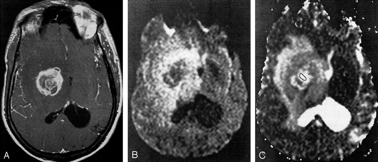
Axial images in an AIDS patient with toxoplasmosis.
A, Axial T1-weighted gadolinium-enhanced MR image. The lesion in the right basal ganglia has an irregular, enhancing rim.
B, DW image (b = 1000 s/mm2). The core of the lesion demonstrates unrestricted diffusion.
C, ADC map of a toxoplasmosis lesion in the right basal ganglia. The outlineindicates the ROI within the lesion used for ADC computation. The core of the lesion has a mean ADC that is increased relative to that in normal white matter (ADC ratio, 2.23).
症例 AIDS患者における悪性リンパ腫
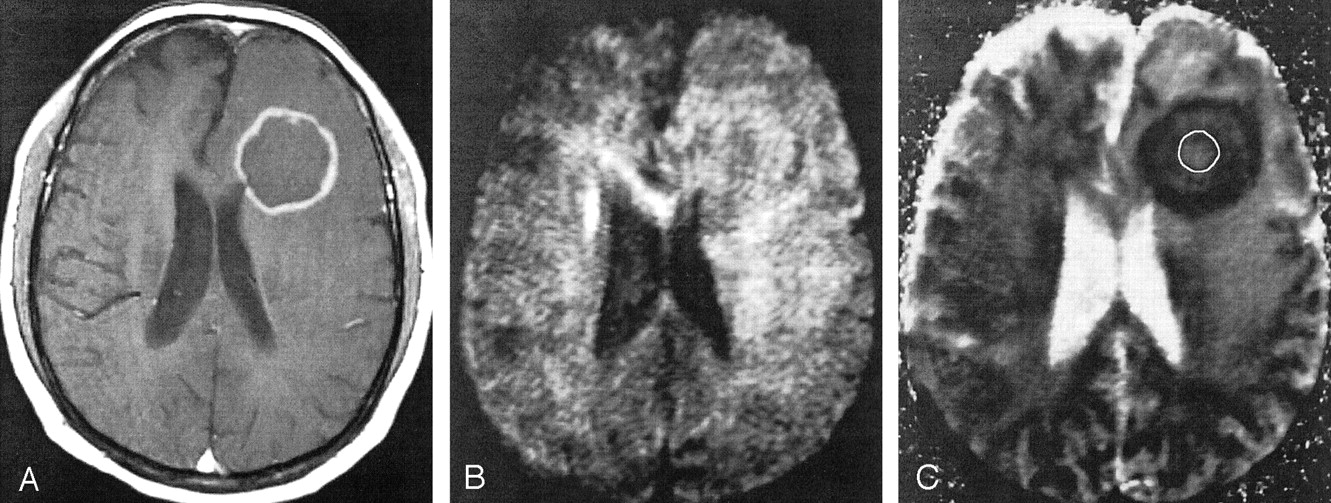
Axial images in an AIDS patient with lymphoma.
A, Axial T1-weighted gadolinium-enhanced MR image. A lesion with an enhancing rim is present in the left frontal lobe.
B, DW image (b = 1000 s/mm2). The signal intensity of the core of the lesion is similar to that of uninvolved white matter.
C, ADC map of a lymphoma lesion in the left frontal lobe. The outline indicates the ROI within the lesion used for ADC computation. The core of the lesion has a mean ADC that is similar to that of normal white matter (ADC ratio, 1.25).
2症例ともAJNR 24:633-637,2003より引用
よくある疑問(Q&A)
Q. トキソプラズマに感染している人は皆、脳症になりますか?
A. なりません。多くは体内に潜んだまま過ごし、脳症は免疫が大きく低下したときに再活性化して起こります。免疫の病気や治療中の方で新しい神経症状が出たら、早めに主治医に相談してください。
参考文献
- Camacho DL, Smith JK, Castillo M. Differentiation of toxoplasmosis and lymphoma in AIDS patients by using apparent diffusion coefficients. AJNR Am J Neuroradiol. 2003;24(4):633-7. PubMed
- Kumar GG, Mahadevan A, Guruprasad AS, et al. Eccentric target sign in cerebral toxoplasmosis: neuropathological correlate to the imaging feature. J Magn Reson Imaging. 2010;31(6):1469-72. PubMed
- Sakai M, Higashi M, Fujiwara T, et al. MRI imaging features of HIV-related central nervous system diseases: diagnosis by pattern recognition in daily practice. Jpn J Radiol. 2021;39(11):1023-1038. PubMed
- Elicer I. Approach to an Intracranial Mass in Patients With HIV. Curr Neurol Neurosci Rep. 2020;20(9):43. PubMed
- Dunlop O, Rootwelt V, Sannes M, et al. Risk of toxoplasmic encephalitis in AIDS patients: indications for prophylaxis. Scand J Infect Dis. 1996;28(1):71-3. PubMed
- Luft BJ, Hafner R, Korzun AH, et al. Toxoplasmic encephalitis in patients with the acquired immunodeficiency syndrome. Members of the ACTG 077p/ANRS 009 Study Team. N Engl J Med. 1993;329(14):995-1000. PubMed
- Lewitschnig S, Gedela K, Toby M, et al. 18F-FDG PET/CT in HIV-related central nervous system pathology. Eur J Nucl Med Mol Imaging. 2013;40(9):1420-7. PubMed
- Pomeroy C, Filice GA. Pulmonary toxoplasmosis: a review. Clin Infect Dis. 1992;14(4):863-70. PubMed
画像診断を無料で学べるコンテンツ

腹部画像診断を症例で学ぶ
4日に1回、朝6時に症例が届き、実際に画像をスクロールして読影する講座です。90症例以上・1年以上続く講座で、医師・医学生・放射線技師・看護師など10,000名以上が参加中です。
講座に参加する(無料) →