トキソプラズマ脳症とは?
- AIDS指標疾患の1つ。AIDS患者ではCD4<100/μLで発症する。
- 潜伏感染のトキソプラズマ原虫の再活性により発症する。
- 脳炎、肺炎、心筋炎などを合併することがある。
- 脳では、悪性リンパ腫との鑑別が問題となる。(なぜならばAIDSに合併するリンパ腫はリング状造影効果を呈するため)
- ピリメタミン(抗原虫薬)、サルファ剤(抗菌薬)などを投与して臨床的あるいは画像上に改善があれば、トキソプラズマ、なければ悪性リンパ腫という診断的治療を行なうこともある。
トキソプラズマ脳症の画像所見
- 基底核に好発する。
- 周囲に浮腫を伴う単発もしくは多発腫瘤。
- 結節状もしくはリング状に造影される。
- リング状の濃染内に偏在性の結節状濃染(eccentric target sign:2割)
- DWIにて等信号。
- 悪性リンパ腫とは、ADC比が1.6以上の場合はトキソプラズマと診断できるが、しかし、ADCではoverlapも多い。→FDG-PETが有効という報告もある。
- 他、201Tl-SPECTが鑑別に有用との報告もある。集積が見られれば悪性リンパ腫の可能性が高い。しかし、どれも微妙。→診断的治療を行なうことあり。
症例 AIDS患者におけるトキソプラズマ脳症
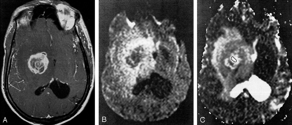
Axial images in an AIDS patient with toxoplasmosis.
A, Axial T1-weighted gadolinium-enhanced MR image. The lesion in the right basal ganglia has an irregular, enhancing rim.
B, DW image (b = 1000 s/mm2). The core of the lesion demonstrates unrestricted diffusion.
C, ADC map of a toxoplasmosis lesion in the right basal ganglia. The outlineindicates the ROI within the lesion used for ADC computation. The core of the lesion has a mean ADC that is increased relative to that in normal white matter (ADC ratio, 2.23).
症例 AIDS患者における悪性リンパ腫
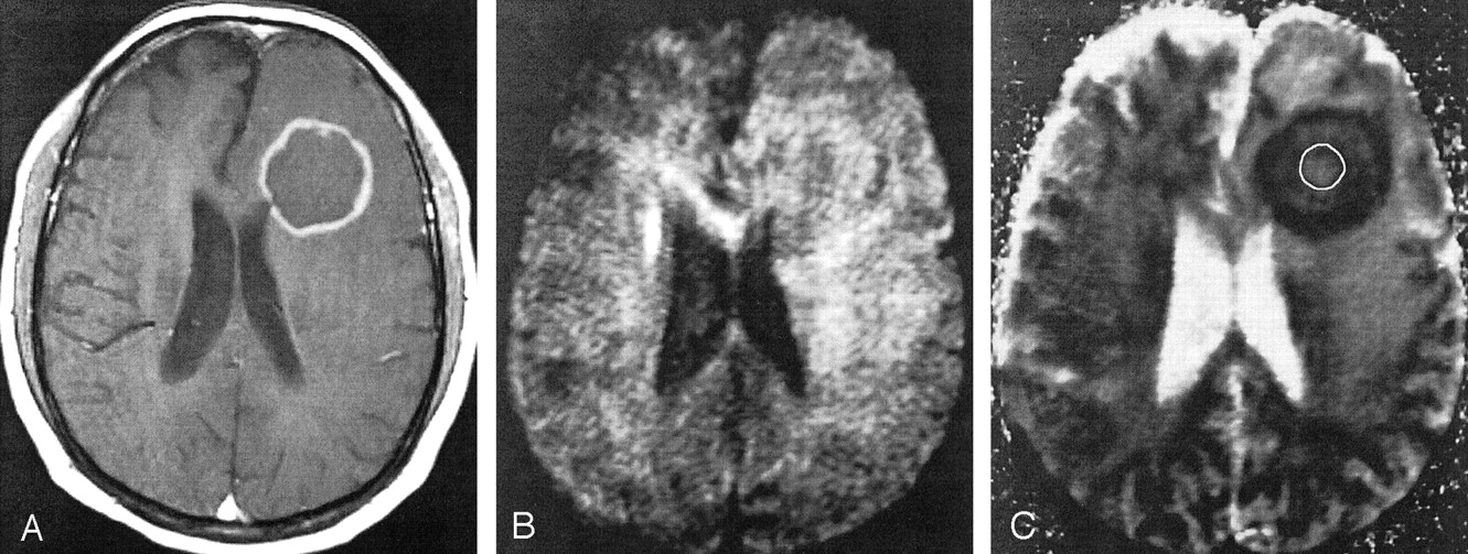
Axial images in an AIDS patient with lymphoma.
A, Axial T1-weighted gadolinium-enhanced MR image. A lesion with an enhancing rim is present in the left frontal lobe.
B, DW image (b = 1000 s/mm2). The signal intensity of the core of the lesion is similar to that of uninvolved white matter.
C, ADC map of a lymphoma lesion in the left frontal lobe. The outline indicates the ROI within the lesion used for ADC computation. The core of the lesion has a mean ADC that is similar to that of normal white matter (ADC ratio, 1.25).
2症例ともAJNR 24:633-637,2003より引用
ご案内
スマホ版画像診断cafe100日チャレンジ
2026年7月1日から100日で完成を目指します。制作の進捗はメルマガでもお届けします。

腹部画像診断を学べる無料コンテンツ
4日に1日朝6時に症例が配信され、画像を実際にスクロールして読影していただく講座です。現状無料公開しています。90症例以上あり、無料なのに1年以上続く講座です。10,000名以上の医師、医学生、放射線技師、看護師などが参加中。
胸部レントゲンの正常解剖を学べる無料コンテンツ
1日3分全31日でこそっと胸部レントゲンの正常解剖の基礎を学んでいただく参加型無料講座です。全日程で簡単な動画解説付きです。
画像診断LINE公式アカウント
画像診断cafeのLINE公式アカウントで新しい企画やモニター募集などの告知を行っています。 登録していただくと特典として、脳の血管支配域のミニ講座の無料でご参加いただけます。